
How Smart Wearables Work: ECG, PPG, HRV & Sensor Technology Explained
- Farraz Mir
- Apr 1
- 11 min read
Updated: Jun 30
Smart wearable devices turn tiny, continuous body signals into health insights. Technologies like ECG and PPG allow wearables to track heart activity, while metrics like Heart Rate Variability (HRV) and Pulse Rate Variability (PRV) provide deeper insights into stress, recovery, and autonomic function. This article explains how wearable sensors work, the difference between ECG and PPG, how HRV is derived, and why sensor placement and signal processing significantly impact accuracy.
Smart wearable devices started as simple gadgets and now sit at the intersection of bioscience and consumer electronics. What began as basic fitness trackers has now become a category of devices capable of continuously measuring physiological signals and translating them into meaningful health insights. These devices measure parameters such as heart rate, sleep patterns, activity levels and more. But more importantly, they attempt to infer deeper physiological states like stress, recovery, and overall cardiovascular health.
To understand how wearables achieve this, we need to look beyond the interface and into the core sensing technologies that power them.
What is a Smart Wearable Device?
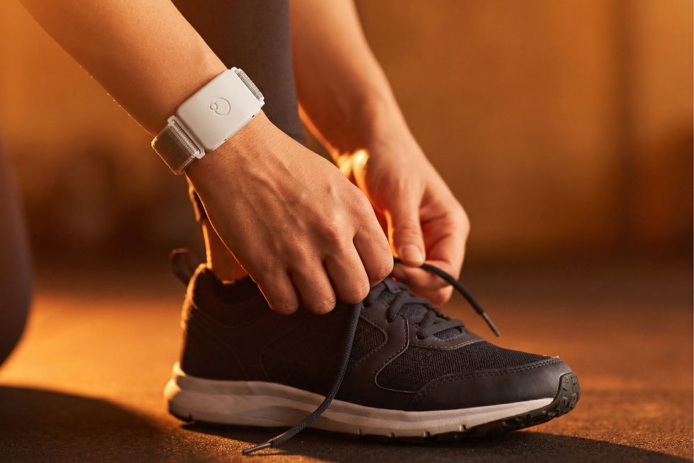
A smart wearable is essentially an IoT (Internet of Things) device embedded with sensors to collect physiological or behavioral data in real time. These devices typically monitor the body continuously, process signals locally or via cloud systems and provide feedback through apps or dashboards.
In medical contexts these devices provide health insights like heart rate, sleep, calories burned. In consumer contexts they support activity and wellness. At their core, however, all wearables rely on one fundamental principle: measuring signals from the human body and converting them into usable data.
A Quick History: Early Wearables to the First Wearable Computer
If we were to expand the definition of wearable devices beyond just IoT or electronic systems, and instead consider any tool worn on the body that enhances human capability - the history of wearables stretches much further back than we might expect.
Early examples include eyeglasses¹, first developed in the late 13th century, which remain one of the most brilliant pieces of biomedical technology ever created. Similarly, the invention of the wristwatch transformed how humans interact with time, making it portable and personal. Even objects like the abacus ring² from the 17th century, or something as fundamental as footwear, which dates back over 8400 years, can be viewed as early forms of wearable technology designed to augment human ability.
However, the earliest known modern form of a smart wearable device was a small computer that could fit into a shoe. Created by Edward Thorp and Claude Shannon in 1961, the "Gambler's Shoe"³ was designed to cheat at casinos (which, in my opinion, exemplifies one of the most fascinating human technological impulses). Their success was not surprising, considering it involved two of the brightest minds of their era. It's almost amusing how mathematicians and scientists have often been driven by the challenge of outsmarting systems, especially the casino's. I wouldn't be surprised if there's something in our DNA that pushes us towards this, given the abundance of movies and literature on the subject.

In many ways, this device represents one of the earliest examples of wearable computing as we understand it today: a system that collects inputs, processes them in real time, and assists decision-making. While this is far from a complete history (which would also include developments like hearing aids and early prosthetics), it highlights the broader evolution of wearables: from passive tools to intelligent, data-driven systems.
Before we jump into how modern wearables work, we need to look at one of the most foundational technologies in physiological measurement: the electrocardiogram (ECG).
The first practical ECG machine was developed in 1903 by Willem Einthoven, who later received the Nobel Prize in 1924 for his work.⁴ His invention built on earlier discoveries by Augustus Waller, who recorded the first human electrocardiogram in 1887, and Luigi Galvani, whose work laid the foundation for understanding electrical activity in living organisms. This shift from mechanical and observational tools to precise electrical measurement, marked a turning point in wearable health technology. This is where wearables begin to move from tools to measurement systems.

From ECG to PPG: The Two Ways Wearables “See” the Heart
Modern wearables primarily use two approaches to measure heart activity: electrical measurement (ECG) and optical measurement (PPG). They are related but different in principle and accuracy.
Electrocardiography (ECG): The Electrical Gold Standard
An electrocardiography (ECG) machine is an electronic device that helps us record the way the heart is pumping blood. ECG measures the electrical activity of the heart. Every heartbeat is initiated by an electrical impulse, and ECG captures this signal with high precision.
The SA (Sinoatrial) node in the heart generates an impulse in its cells by polarising its cells with the influx of Na+ and K+ ions using the sodium-potassium (Na+/K+) ATPase pump (which is an active mode of transporting ions across the cell membrane). This physiological process is important because this is what causes approximately 10⁹ billion cardiac muscle cells in the body to get stimulated. This electrical charge then flows down through the heart's conductive tissues, stimulating them simultaneously until it reaches the ventricles, where depolarisation takes place. Then a depolarisation process occurs, involving K+ ion movement, which causes the heart to return to its resting state by relaxing, and the process continues. The very occurrence of this phenomenon generates a heartbeat, and that is what an ECG records.

The figure above represents an ECG wave. Now, if you pay attention to the generation of a heartbeat, you can correlate them to the P, QRS, and T waves of the ECG. The P wave represents the initial electrical signal that starts a heartbeat, showing how the upper heart chambers prepare to contract. In the QRS complex, the Q represents a small dip, the R is the prominent peak indicating the heart’s primary contraction, reflecting ventricular depolarization, and the S is the downward curve that follows, marking the final phase of this process. The T wave follows, representing the heart muscle relaxing and resetting itself for the next heartbeat. Think of it like an electrical signal traveling through the heart, with each wave showing a different stage of the heart's pumping action from initial preparation, to full contraction, to relaxation. This wave pattern is how doctors and medical devices can read or perhaps see what's happening inside your heart by looking at these simple up and down lines on an ECG graph.
In a normal ECG reading, you will notice multiple waves recorded over multiple minutes. Multiple ECG waves mean multiple R waves. The distance between one R wave and another R wave is referred to as the R-R interval. This is important because it is used to calculate one's heart rate. Your heart rate is calculated by dividing 60 by the RR interval in one minute. This is how an electrocardiogram calculates one's heart rate. The R-R interval between successive R waves gives precise timing for HRV calculation. Clinical/medical-grade HRV is derived from ECG because it captures the cardiac electrical waveform (QRS complex) with millisecond precision.
Because ECG directly measures cardiac electrical activity, it is considered the gold standard for heart rate measurement, HRV analysis and arrhythmia detection.However, ECG requires proper electrode contact, specific placement and often more complex hardware. This makes it less practical for continuous, everyday wear in consumer devices.
Photoplethysmography (PPG)
Photoplethysmography (PPG) is an optical technique used by most consumer wearable devices today. It measures changes in blood volume within the microvascular bed of tissue using LEDs and photodetectors. Unlike ECG, PPG does not measure electrical activity; it measures blood flow dynamics. Let me explain how.
A PPG sensor, over the span of one minute, sends out a light emission. Depending on whether it is a reflective or transmissive PPG, the sensor records the reflected light or absorbs the transmitted light through the location where it was placed. The quality of the acquired signal depends on the underlying physiological and optical properties of the tissue, the wavelength of light used, and the sensor's design which helps it record your Pulse Rate variation or PRV for short.

A PPG signal (see image above) produces a waveform that reflects the pulsatile nature of blood flow. The systolic peak represents maximum arterial blood volume during ventricular systole, corresponding to peak cardiac output and maximum blood ejection, influenced by cardiac contractility, stroke volume, peripheral vascular resistance, and arterial compliance. The dicrotic notch, a physiologically critical inflection point, marks aortic valve closure and represents the transition between systole and diastole, indicating potential reflection waves from peripheral vasculature, arterial stiffness, and vascular health markers. Finally, the diastolic peak signifies peripheral blood volume redistribution, reflecting ventricular relaxation and peripheral perfusion, capturing peripheral vascular tone, autonomic nervous system modulation, and microcirculatory dynamics.
Reflective vs. Transmissive PPG: Placement, Wavelengths and SNR
PPG sensors come in two main configurations: Reflective PPG and Transmissive PPG.

Reflective PPG sensors are often used in wrist-based devices and the forehead. Light is emitted and reflected back to the sensor. It is convenient but often prone to noise and motion artifacts. A reflective PPG sensor uses green light with a wavelength of about 500–570 nm. This relatively shorter wavelength provides the perfect penetration to reach deeper blood vessels where significant changes in blood volume occur. Hemoglobin exhibits distinct absorption peaks in the visible spectrum, particularly around green wavelengths (approximately 540-542 nm). This is due to its molecular structure, which makes green light an ideal candidate for this task because of hemoglobin's absorption spectrum and its ability to penetrate deep enough to penetrate the vascular bed.
On the other hand, in a transmissive PPG sensor, light passes through the tissue and is measured on the other side. It has higher signal quality and better accuracy, and is commonly used in finger-based devices and the earlobe. It uses infrared (IR) and red light as its primary light sources, transmitting it through an organ and measuring the amount of light absorbed. These wavelengths are used because they have longer wavelengths, roughly around 650 nm and 700 nm, which allows them to travel further than other wavelengths of light. The amount of light absorbed depends on the volume of blood present in the tissue at any given time. A photodetector (PD) is usually placed on the opposite side, which records the light and calculates pulse rate variability (PRV) based on the difference between transmitted and absorbed light.
There's a small catch, the readings achieved from a PPG sensor are subject to variation based on where it is placed. Sensor placement has a significant impact on signal quality. This signal quality is further influenced by skin thickness, blood perfusion, motion and temperature. This is why two devices using the same sensor technology can produce very different results.
In the case of reflective PPG, the location can significantly impact the measurement, whether on your wrist or finger. This variation is due to several factors: skin thickness, typical blood flow patterns, skin colour, external light, and the presence of alpha-adrenergic receptors which is a G-protein coupled receptor that plays a role in vasodilation through specific cellular pathways, thereby affecting blood flow and consequently the sensor readings. The finger, earlobe, and ventral (lower) side of the body are considered to have the best identifiable waveform characteristics and a higher Signal-to-Noise (SNR) ratio. The SNR is a critical metric in evaluating PPG sensor performance, as it quantifies the level of desired signal relative to background noise. Surprisingly, the forehead is also an excellent location, while the dorsal (upper) side of the wrist is the worst for recording PPG measurements.
HRV vs PRV: What’s the Difference - and why it Matters
One of the most misunderstood aspects of wearable health tracking is the difference between HRV and PRV. At a surface level, both seem to measure the same thing: how your heart behaves over time. But the way they are captured, and what they truly represent, are fundamentally different.
HRV vs PRV: The Core Difference
Heart rate variability (HRV) is derived from ECG. It measures the variation in electrical heartbeat intervals (R-R intervals). It is considered the gold standard for autonomic nervous system analysis. Pulse rate variability (PRV) is derived from PPG. It measures the variation in pulse arrival times and is influenced by vascular and hemodynamic factors. While PRV often correlates with HRV under ideal conditions, they are not identical.
Now, this is where things get slightly misleading, especially in the world of consumer wearables. Technically, HRV can only be calculated using an ECG because it relies on the QRS complex, specifically the R–R interval, i.e. the time between successive electrical heartbeats). A PPG sensor, on the other hand, does not measure electrical activity. It measures blood flow. Which means it cannot truly calculate HRV.
Instead, what most wearables calculate is PRV: the interval between successive pulse peaks in the optical waveform. Each peak corresponds to a heartbeat, similar in concept to R–R intervals, but fundamentally different in origin. So when many devices say they are showing “HRV,” they are often presenting a PRV-derived approximation. While PRV is a useful surrogate in many scenarios, it does not fully capture the same physiological information as HRV, especially under dynamic conditions.
Why This Matters: The Autonomic Nervous System Connection
To understand why HRV is such an important metric, we need to briefly look at the autonomic nervous system (ANS). In theory, our nervous system which is arguably one of the most fascinating systems in the body, is divided into:
The Central Nervous System (CNS) (brain and spinal cord)
The Peripheral Nervous System (PNS) (all the nerves connecting the body)

The PNS further branches into the autonomic nervous system, which is then subdivided into the parasympathetic and sympathetic nervous systems. This is where things start to get interesting and more relevant to HRV and PRV.
The parasympathetic nervous system is essentially the "rest and digest" system. It activates when we are relaxed and safe. At a molecular level, it starts when our brain sends a relaxation signal through the vagus nerve, which releases a chemical messenger called acetylcholine. This messenger acts like a key that fits into the M2 receptor on our heart cells. When these locks are activated, they trigger two main changes in the heart cells: they open channels that let potassium flow out and reduce the amount of calcium entering the cells. As a result, the heart slows down, beats more efficiently, and variability increases. Think of it like easing off the accelerator and gently applying the brakes.
The sympathetic nervous system works as our body's "fight or flight" mechanism through an energising chain of events. It begins when our brain perceives stress or danger and signals through sympathetic nerves, which release a chemical messenger called norepinephrine. At the same time, the adrenal glands release adrenaline into the bloodstream for a stronger, longer-lasting effect. These chemicals bind to two types of receptors in our body: beta-receptors in the heart and alpha-receptors in blood vessels. When norepinephrine binds to beta-receptors in the heart, it triggers a series of changes that increase calcium levels inside heart cells, making the heart beat faster and stronger, much like pressing the accelerator in a car. When it binds to alpha-receptors in blood vessels, it makes them constrict, which helps raise blood pressure. This whole process is our body's way of preparing for action: increasing blood flow to muscles, raising alertness, and getting ready for whatever challenge lies ahead. It's like our body's natural energy boost system, preparing you to either fight or run away from danger.
Heart rate variability is essentially a reflection of how these two systems interact:
Higher HRV =Strong parasympathetic activity (rested, recovered, adaptable)
Lower HRV = Dominant sympathetic activity (stress, fatigue, strain)
Since HRV measures the variation between R–R intervals, a larger variation indicates a healthier, more adaptable system. A smaller variation means a more rigid, stressed system. This is why HRV is widely used as a marker of recovery, stress and overall physiological resilience.
Now here’s the important connection.
PRV tries to estimate this same variability, but through blood flow signals instead of electrical signals. Because of this it introduces additional noise, captures vascular effects along with cardiac activity and may miss subtle autonomic changes that ECG can detect. In simple terms: HRV measures the heart’s electrical rhythm directly. PRV measures the downstream effect of that rhythm through blood flow. They are related, but not interchangeable.
This distinction becomes critical when interpreting wearable data. A smartwatch might show “HRV,” but it is often PRV-based. Trends are still useful, but absolute values may not be clinically equivalent. However, this doesn’t make wearables useless - far from it actually. What it simply means is that they should be used as directional tools, not diagnostic instruments.
Conclusion
Smart wearable devices are powerful tools that translate biological signals into everyday insights. By combining sensor technologies like ECG and PPG with advanced signal processing, they allow continuous monitoring of the human body in ways that were previously only possible in clinical settings.
However, understanding what these devices actually measure, and their limitations, is essential. Metrics like HRV and PRV provide valuable insights, but their accuracy depends on sensor design, placement, and algorithmic interpretation. As technology advances, wearables will play an increasingly important role in preventive healthcare. But for now, the key lies in using them not as absolute diagnostic tools, but as directional indicators of physiological trends.
References:
Medievalists.net. (2022, March 16). Medieval eyeglasses: Wearable Technology of the thirteenth century. https://www.medievalists.net/2016/03/medieval-eyeglasses-wearable-technology-of-the-thirteenth-century/
Wearable abacus from the Qing Dynasty. List of Physical Visualizations. (2016, July 16). https://dataphys.org/list/wearable-abacus-from-the-qing-dynasty/
Ed Thorp, Claude Shannon and the world’s First Wearable Computer. Winton. (n.d.). https://www.winton.com/news/ed-thorp-claude-shannon-and-the-worlds-first-wearable-computer
Wikimedia Foundation. (2026, March 8). Willem Einthoven. Wikipedia. https://en.wikipedia.org/wiki/Willem_Einthoven


Comments